


Online Dental Trauma Guide: Part I Fracture Injuries
Alveolar fractures
Alveolar fractures
Fractures involving the alveolar bone which may extend across several teeth.
Clinical findings
The common presentation of an alveolar fracture is:
Segment mobility with several teeth moving en-bloc
Changes in the occlusion (due to misalignment of the fractured alveolar segment)
Often associated with luxation injuries, notably lateral luxations and avulsions.
Radiographic findings
Multiple projection views, including a panoramic radiograph can:
Be helpful in determining the course and position of the fracture
Present as vertical or horizontal radiolucent lines in between or over the roots of the teeth
Be located at any level from the marginal bone to the root apex.
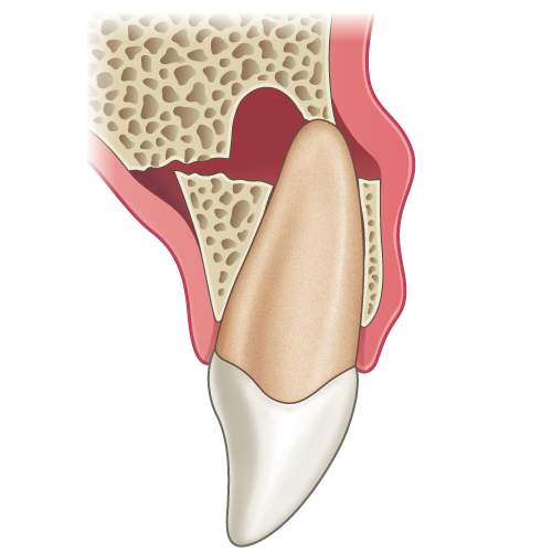
Illustration: Longitudinal section of a tooth showing an alveolar fracture.

Alveolar fracture in the region of the apices of the lower incisors.
Management
Primary teeth
The displaced segment should be digitally repositioned under local or general anaesthesia and flexibly splinted for 4 weeks.
Secondary teeth
The displaced segment should be digitally repositioned under local or general anaesthesia and flexibly splinted for 4 weeks.
Monitoring: For primary teeth follow-up is advised until exfoliation of the tooth. For permanent teeth the follow-up period advised is yearly for 5 years.