


Subluxation
Subluxation
Subluxation is a moderate injury to the tooth-supporting structures.
Clinical findings
A subluxated tooth will be:
Tender to touch
Mobile
In the normal position with no change in the occlusion
Associated with bleeding from the gingival crevice (this is a pathognomonic sign and due to cleavage and tearing of the periodontal ligament).

Illustration: Longitudinal section of a tooth showing a subluxation injury.
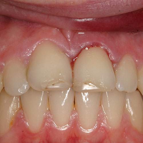
Clinical image: Labial view showing bleeding from gingival margin of subluxed UL1.
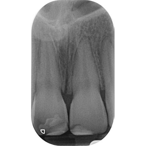
Radiograph: Subluxed UL1 showing normal periodontal ligament space.
Radiographic findings
Of a subluxated tooth will show:
A healthy-looking tooth
No obvious widening or changes in the periodontal ligament.
Management of primary teeth
Reassure the patient (and their parents/ carers)
Monitoring is advised
If the tooth is very mobile (due to a resorbed root being close to exfoliation) and poses a potential airway risk – the tooth should be removed.
Management of permanent teeth
Reassure the patient
Advise a soft diet for 2 weeks
Encourage patients to brush their teeth normally as soon as possible
Tooth discolouration soon after the injury may be transient. Patients should therefore be reassured and no diagnosis of necrosis made in the absence of any other signs or symptoms (particularly within the first three months after the injury)
Monitor the pulp for 1 year
Follow-up thereafter is as per the scheduled review for that patient.
Splinting
IS IT NECESSARY?
In some situations owing to the heightened emotional state of the patient, splinting subluxed teeth can help reassure patients, particularly if the tooth is very tender and it may also protect the apex.
SPLINTING TIME
A flexible splint for 2 weeks maybe helpful.
WHAT MATERIALS TO USE?
Several different materials are available for splinting a 0.016″ stainless steel orthodontic wire with composite resin.
This should be extended to one uninjured tooth either side of the injured teeth.
Monitoring: The pulpal response should be monitored at subsequent appointments until a definitive pulpal diagnosis can be made and a follow-up period of one year is advised.